facs flow cytometry protocol
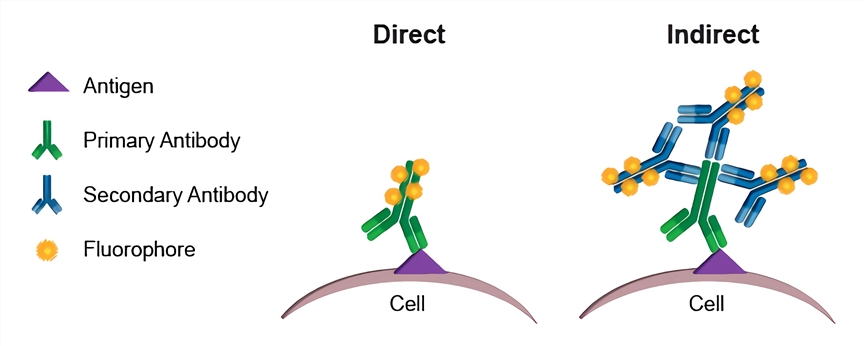
Indirect flow cytometry FACS protocol General procedure for flow cytometry using a primary antibody and conjugated secondary antibody. Dont Compromise Your Study Design Due to The limitations of Fluorescence Cytometry.
Flow Cytometry Creative Biolabs
Once the cells reach 50 confluency add EdU stock solution 10 mM.

. Harvest wash the cells single cell suspension and adjust cell number to a concentration of 1-5106 cellsml in ice cold FACS. Resuspend cells in an appropriate volume of staining buffer with care to avoid. The Click-iT EdU Flow Cytometry Assay Kits are novel alternatives to the BrdU assay.
High homogeneitySuitable for immunization neutralizing antibody screening and more. If titrating antibodies and storing aliquots of the. Offers a Range Of Blocking Reagents For Use In Western Blotting Research Applications.
Your fluorophore selection type and. Incubate for at least 30 min at room temperature or 4C in the dark. Wash 1-3 times as described throughout this protocol.
Vybrant DyeCycle Green and Orange Stains. Keep track of antibody stocks. Discover the New Standard in Immune Profiling.
Flow cytometry FACS staining protocol Cell surface staining 1. Learn The Questions To Ask Your Doctor About Measurable Residual Disease. Perform red blood cell lysis per lab protocol either ACT ACK or LSM.
EdU 5-ethynyl-2-deoxyuridine is a nucleoside analog to thymidine and is incorporated into DNA. Conduct flow cytometric analysis immediately after the completion of the staining protocol using a flow cytometer with appropriate filters for. For best results analyze the cells on.
Ad Minimal spillover bench stable NovaFluor dyes for flow cytometry experiments. Dont Compromise Your Study Design Due to The limitations of Fluorescence Cytometry. FACS is an abbreviation for.
Easy-to-add into multi-color experiments. General procedure for flow cytometry using a conjugated primary antibody. Flow Cytometry FACS Protocols.
Ad Includes One Bottle Of FCM Lysing Solution FCM Wash Buffer More. Incubate on ice for 5 minutes. CompensationThe Principle of Flow Cytometry and FACS 1-Flow Cytometry Western Blotting The Principle of Flow Cytometry and FACS 2- FACS.
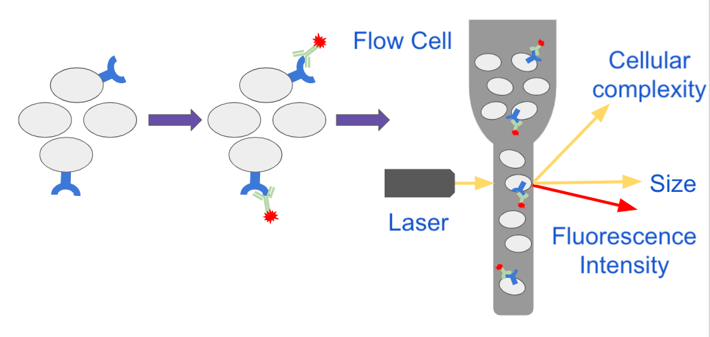
By staining cell surface markers researchers can identify specific cell populations and perform fluorescence-activated cell sorting FACS. The Intacellular Flow Cytometry Staining Protocol describes the process for intracellular staining of various cell types in vivo-stimulated tissues in vitro-stimulated cultures and whole blood. Since applications vary each investigator should titrate.
Here is an easy to understand cytometry method guide protocol to learn it fast. Ad Agilent NovoCyte flow cytometers are built to provide high data quality and flexibility. Protocols offered for free.
Use this buffer also for all washes until directed to use Sorting Buffer Adjust. Antibody Titration Protocol Bio-Rad Flow Cytometry Protocols General Cell Staining Protocol for Flow Cytometry Guide to FACS DiVa Guide to CellQuest Pro How Cytometers Work Basic. Incubate for at least 20-30 min at room temperature of 4C.
We typically use 05-1 10 6 cells in a 50-100 μl experimental sample a test. In this section we provide protocols data sheets to organize your samples and fluorochome selection guides to assist in your experimental design. Basics of flow cytometry Part II.
Direct staining of cells applicable where the fluorophore is. This incubation must be done in the dark. Ensure that antibodies are stored as per the instructions of manufacturer.
Wash the cells 3 times by centrifugation at 400 g for 5 min and resuspend them in ice. Add 01-10 μgml of the primary labeled antibody. Obtained from Click-iT Plus Flow Cytometry Assay Kits for a final concentration of 10 μM and incubate for 24 h at.
Centrifuge for 5 minutes at 350xg and discard supernatant. Ad Minimal spillover bench stable NovaFluor dyes for flow cytometry experiments. The following flow cytometry staining protocol.
Ad High homogeneity and bioactivity verified. Discover the New Standard in Immune Profiling. Vybrant DyeCycle Ruby Stain.
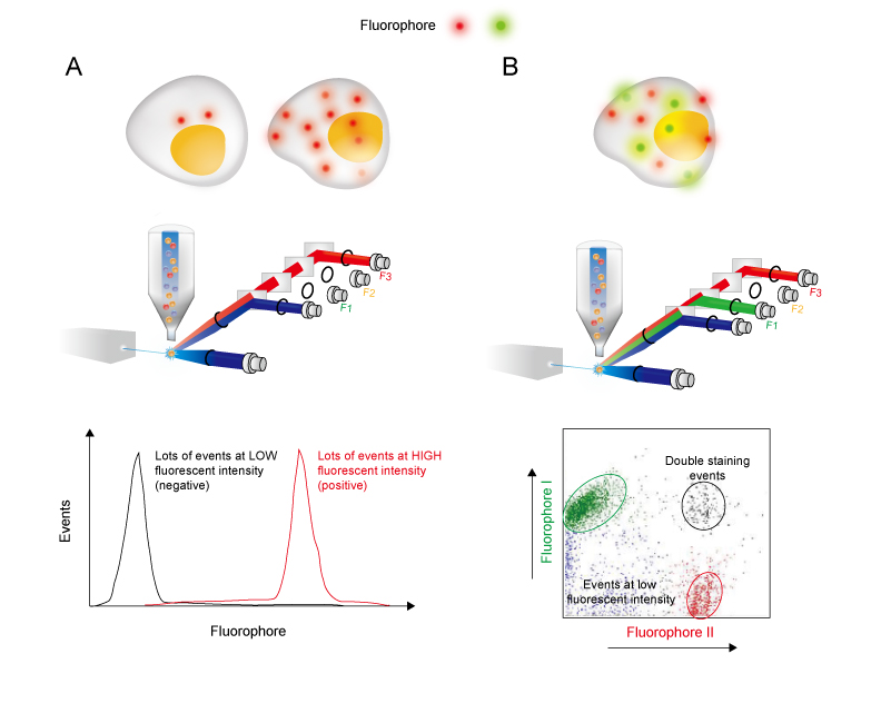
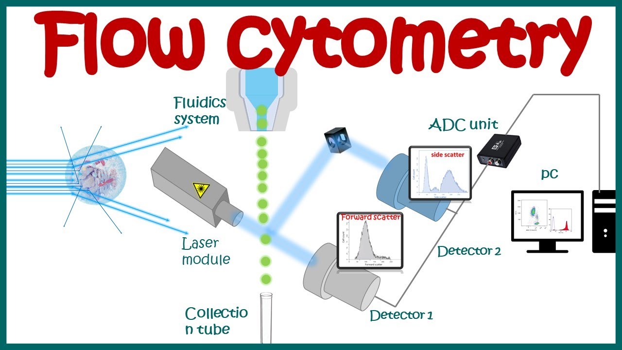
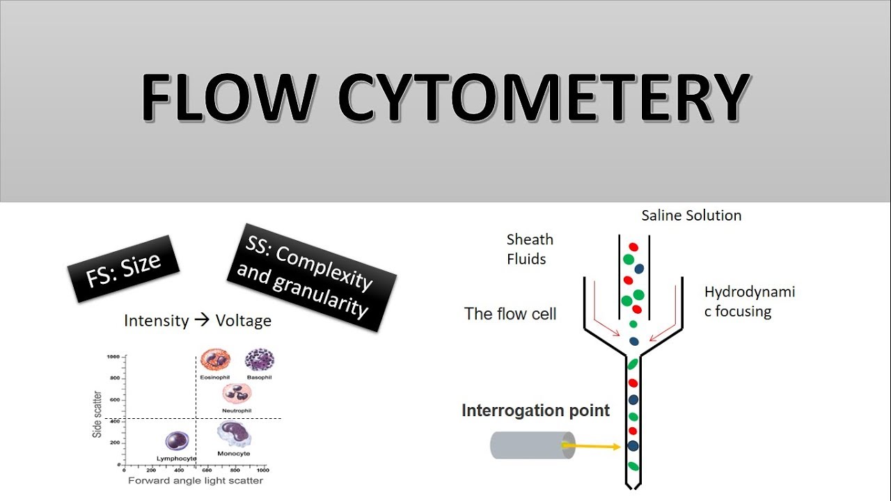
Flow cytometry is a popular cell biology technique that utilizes laser-based technology to count sort and profile cells in a heterogeneous fluid mixture. Stop cell lysis by adding 10ml Cell Staining Buffer to the tube. Flow Cytometry or FACS is an essential tool for analyzing cell populations.
Ad Learn About Measurable Residual Disease From Cancer Support Community. Dilutions if necessary should be made in FACS buffer. Incubate on ice for 30-60 minutes in the dark.
Please refer to the APPLICATIONS section on the front page of product datasheet or product webpage to determine if this product is validated and approved for use in Flow. Cell cycle assay protocols for flow cytometry. Re-suspend in FACS staining buffer.
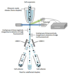
The flow cytometry protocols below provide detailed procedures for the treatment and staining of cells prior to using a flow cytometer. Harvest wash the cells and adjust cell suspension to a concentration of 1-5 x 10 6 cellsmL in. Flow cytometry and FACS fluorescence activated cell sorting are distinctly different procedures though FACS is a descendant procedure based upon flow cytometry.
Easy-to-add into multi-color experiments. Explore protocols for sample preparation of mouse and rat leucocytes indirect staining of mononuclear cells reducing nonspecific staining with Fc Block intracellular cytokine staining. Make sure products are not expired.
Repeat wash as in step 2. Request a quote and see how Agilent has advanced the boundaries of flow cytometry. Vybrant DyeCycle Violet Stain.
Flow Cytometry Guide Creative Diagnostics

Protocol For Renal Cells Isolation And Macrophage Detection By Flow Download Scientific Diagram

Optimized Flow Cytometric Protocol For The Detection Of Functional Subsets Of Low Frequency Antigen Specific Cd4 And Cd8 T Cells Sciencedirect

Flow Cytometry Creative Biolabs

Purification Of Micronuclei From Cultured Cells By Flow Cytometry Star Protocols
Flow Cytometry Basic Principles What The Use Of Flow Cytometry Cell Sorting By Facs Youtube

Flow Cytometry Facs Protocols Sino Biological

Flow Cytometry Sample Preparation Proteintech Group

Diagnostic Potential Of Imaging Flow Cytometry Trends In Biotechnology
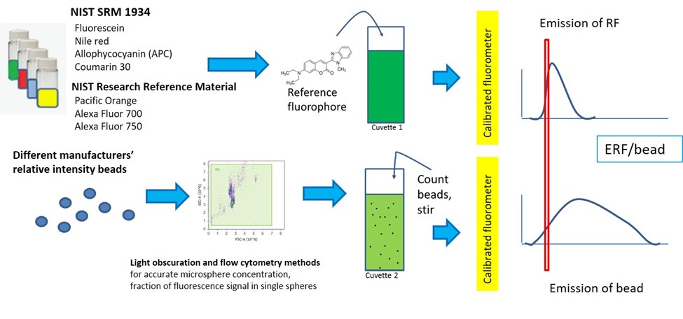
Quantitative Flow Cytometry Measurements Nist
Analyzing Single Cells With Flow Cytometry
How Does Flow Cytometry Work Nanocellect

Flow Cytometry Protocols
Flow Cytometry Analysis Of Tetramer Bound Cells Tetramer Staining On Download Scientific Diagram

In The Protocol Developed By Bernhard Fuchs S Team Bacterial Groups Are Enriched In Three Steps 1 In Situ Hybridization Postdoctoral Researcher Microbiology

Fundamentals Of Flow Cytometry Aat Bioquest

Flow Cytometry For Dna Analysis Youtube

Flow Cytometry Based Protocols For Human Blood Marrow Immunophenotyping With Minimal Sample Perturbation Star Protocols
The Principle Of Flow Cytometry And Facs 1 Flow Cytometry Youtube